
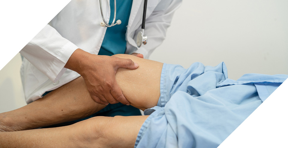
Cancer Procedures
Interventional radiology offers targeted and minimally invasive techniques for precise cancer procedure and care.
Offerings:
Chemoembolization
Chemoembolization is a minimally invasive procedure that treats certain types of cancer, particularly liver cancer. It uses a combination of chemotherapy drugs and blood vessel embolization to block a tumor's blood supply. Without the blood supply to feed the tumor, it begins to shrink and die.
What you need to know
During chemoembolization, an interventional radiologist will insert a catheter into the artery, guide it to the blood vessels that supply the cancerous tumor, and inject contrast dye for real time visualization purposes. From there, a mix of chemotherapy drugs that attack the cancer cells directly, and an embolic agent that blocks blood flow and prevents further growth, is injected.
This procedure requires only local anesthesia and IV sedation and lasts one to two hours – not including preparation and recovery time. Patients may be required to spend the night at the hospital for observation and pain medications or may be released the same day.
The location and size of the tumor, as well as a patient's overall health, are also important factors in determining whether chemoembolization is the right course of procedure. Certain health conditions, such as blood clotting disorders or kidney disease, may prohibit your ability to undergo chemoembolization.
For some, chemoembolization is the only procedure necessary. For others, it may be used in conjunction with surgery, systemic chemotherapy, ablation, or radiation therapy.
Schedule an appointment?
LET'S GET STARTEDPleurX
Lungs are surrounded by two layers of tissue called pleura. Within this pleural space there is fluid to assist with smooth breathing function. When too much fluid accumulates in this area, it can make it difficult to breathe. Similarly, fluid buildup in the abdomen and peritoneal cavity, also known as ascites, can have painful symptoms and serious effects on surrounding organs.
The PleurX drainage catheter is a thin, flexible tube placed in the chest to drain excess fluid from around the lungs or abdomen.

What you need to know
Using real time imaging technology, the PleurX is guided into the lung's pleural space or abdomen's peritoneal cavity. Once in place, the catheter is secured to the skin with a suture or adhesive dressing. The other end of the tube has a one-way valve and connector that allows for the attachment of a drainage system that consists of a vacuum or manual pump to draw fluid out, and a bottle or bag to collect it.
This procedure makes it possible to drain excess fluid at home. Following instructions (regarding frequency and duration) and training given by the overseeing healthcare provider, the patient or caregiver can connect the drainage system to the catheter and open the one-way valve to drain excess fluid out of the body and into the collection bag or bottle.
The PleurX system can help alleviate uncomfortable or painful symptoms and improve the quality of life for patients with recurrent pleural effusions or malignant ascites. As an added benefit, the end of the PleurX system catheter is covered by a thin dressing and can be discreetly hidden beneath your clothing.
Schedule an appointment?
LET'S GET STARTEDPorts
Implanted under the chest skin, a port provides a safe and reliable way to inject medicine and take blood samples. This device is connected to a catheter that provides easier and less painful access for those who need frequent intravenous treatments, transfusions, or blood draws.
What you need to know
A port implantation is an outpatient procedure performed by an interventional radiologist while the patient is under light anesthesia. A small incision is made in the neck to access the vein using imaging as guidance. Another small incision is made in the chest, arm, or abdomen for port placement. After connecting the two via catheter, a pouch is created beneath the skin and the port is implanted.
An X-Ray is performed to ensure the port is in the correct location and the incisions are closed with dissolvable stitches or surgical adhesive. Recovery time for this procedure is relatively short and a slight bulge in the skin where the port was placed may be visible.
Going forward, needles can be inserted into the port's silicone top or septum. This is followed by another needle that flushes the device to prevent blockages or blood clots. The site is then covered with a clear bandage to ensure symptoms of infection (like red streaks) are visible.
- Are receiving ongoing chemotherapy.
- Are undergoing long-term intravenous therapy.
- Have poor vein access.
- Receive frequent blood transfusions.
- Require regular blood sampling.
Schedule an appointment?
LET'S GET STARTEDTumor Ablation
When surgical removal of a tumor is not feasible, a minimally invasive tumor ablation procedure may be used to shrink or destroy the tumor. Using thermal energy, high frequency electrical currents, or freezing methods, the tumor is selectively reduced or eliminated with minimal damage to the surrounding tissues.

What you need to know
- Radiofrequency Ablation: During this procedure, a needle-like electrode is inserted into the tumor under imaging guidance. Once properly positioned, high frequency electrical currents are passed through it, generating heat. The heat creates friction within the tumor, which leads to the death of the tumor cells.
- Microwave Ablation: This procedure also uses a needle-like probe that is guided into the tumor using imaging guidance. Instead of electrical currents, microwave energy is delivered through the probe. This generates heat and effectively kills the tumor cells.
- Cryoablation: During this procedure, tumor tissue is destroyed through a freezing process. Cryoprobes are thin, hollow, and needle-like probes, through which extremely cold gases or liquids can flow. This process cools the surrounding tissue and ice crystals form within the freezing cells. Direct freezing occurs and the blood supply to the tumor is cut, which leads to cell death of abnormal or diseased tissue.
Talk to your healthcare provider today to determine if tumor ablation is the right option for you.
Often used in conjunction with other procedure methods, like chemotherapy or surgery, tumor ablation can significantly improve overall patient outcomes.
Schedule an appointment?
LET'S GET STARTEDY90
Y90, also known as yttrium-90 radioembolization, is a safe and highly effective internal radiation therapy used to treat liver cancer. During this outpatient procedure, tiny, radioactive beads known as microspheres are injected into the blood vessels that supply tumors in the liver to shrink them and improve liver function.

What you need to know
Once the blood vessels feeding the tumor are identified, the Inerventional Radiologist will map out the procedure plan to determine appropriate dosage and activity level of the Y90 microspheres, small radioactive particles, to be administered.
During the procedure, the Y90 microspheres are injected through a catheter into the target blood vessels, lodging themselves into the tumor, and releasing a high dose of radiation to kill cancer cells.
After their injection, the catheter is removed, and pressure is applied to the insertion site to minimize bleeding. Following a few hours of observation, the patient is discharged.
The decision to undergo Y90 procedure will be made in conjunction with a healthcare provider that can evaluate your individual needs. Talk to your doctor today to find out if Y90 is right for you.
Schedule an appointment?
LET'S GET STARTEDCare that covers Kansas City communities in Kansas & Missouri.
The Clarity Care Imaging & Specialty Care office is conveniently located at 9040 Quivira Road in Lenexa, Kansas. Our center is just north of Oak Park Mall with easy access from much of the Kansas City area including Overland Park, Olathe, Shawnee, Merriam, Mission, Leawood and Prairie Village.