Venous Disease
Interventional radiology offers targeted and minimally invasive techniques for precise venous disease procedure and care.
Offerings:
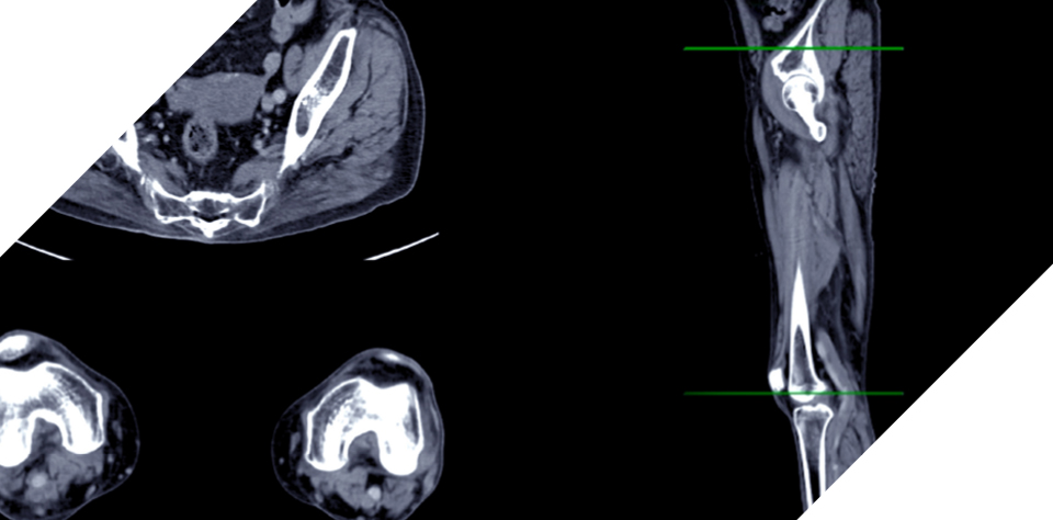
Extremity Venogram
An extremity venogram is a diagnostic imaging procedure used to visualize the veins in the arms or legs. It is commonly performed to detect and evaluate conditions such as deep vein thrombosis (DVT), venous insufficiency, or venous malformations.
What you need to know
During an extremity venogram, an iodine-based contrast dye that can be seen on X-rays is injected into a vein, usually in the foot or hand. From there, a series of X-Ray images is taken to visualize the veins and any abnormalities, such as blood clots or blockages. During the imaging process, the patient may be asked to assume different positions, such as lying flat or elevating the extremity being examined. Compression may also be applied to assess venous flow and detect abnormalities.
Following the procedure, a highly trained Clarity Care radiologist reviews and interprets the images to evaluate the structure and function of the veins and develop a course of procedure. Patients can resume normal activities with little to no down time.
If you are experiencing symptoms or have risk factors for venous conditions such as:
- Deep vein thrombosis (DVT), which occurs when a blood clot forms in a deep vein, usually the leg.
- Venous insufficiency, which occurs when valves in the veins of the legs or arms do not function properly, leading to blood pooling and venous congestion.
- Venous malformations, which are abnormalities within the veins, such as tumors or congenital malformations.
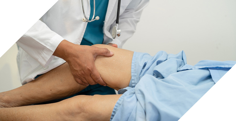
- Chronic leg swelling or pain, which can be assessed to determine underlying causes.
To learn more, schedule a consultation with one of Clarity Care's skilled interventional radiologists to evaluate your specific condition, review your medical history, and consult imaging results.
Extremity venograms are extremely effective tools for diagnosing and evaluating venous conditions in the arms and legs. Because venograms provide detailed images of the veins, they allow healthcare providers to visualize structure, blood flow, and abnormalities within the veins.
Extremity venograms are a low-risk procedure. There is a low risk of an allergic reaction to the contrast dye. There is some risk of damage to the kidneys in individuals with existing kidney conditions and low radiation exposure due to X-ray technology.
It is important for patients to discuss concerns or potential risks with their healthcare provider to determine if the benefits outweigh potential drawbacks.
Schedule an appointment?
LET'S GET STARTEDMay-Thurner Syndrome
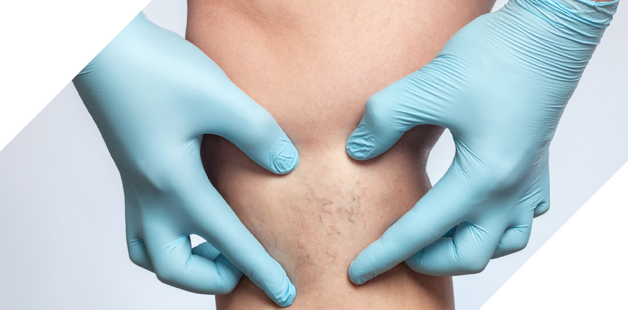
May-Thurner syndrome, also known as iliac vein compression syndrome, is a vascular condition characterized by compression or the narrowing of the left common iliac vein, a major blood vessel located in the pelvis. The primary function of this vein is to return blood from the lower limbs and pelvis back to the heart. When compressed, it can lead to reduced blood flow, venous stasis, and the formation of blood clots.
What you need to know
The procedure for May-Thurner syndrome aims to relieve compression, improve blood flow, and prevent the formation of blood clots. The most common procedure options include:
- Anticoagulation: Medications such as heparin and warfarin may be prescribed to prevent the formation of blood clots by thinning the blood.
- Thrombolytic Therapy or thrombectomy. In cases where blood clots have already formed, thrombolytic therapy or a clot removal device may be used. This involves the administration of medications or removal of the clot to restore normal blood flow.
- Angioplasty and Stenting: In cases where the compression of the left iliac common vein is severe and causes significant symptoms, a small balloon is inflated to open the narrowed vein and a stent is placed to relieve compression and restore blood flow.
- Surgical Bypass: If other procedure options are not successful, rarely a surgical bypass may be considered. This involves creating a new pathway for blood flow by surgically connecting the affected vein to a nearby healthy vein.
If you are experiencing mild to severe symptoms associated with May-Thurner syndrome, you may be a candidate for procedure at Clarity Care. Common symptoms include:
- Leg pain, a hallmark symptom of May-Thurner syndrome, is described as a dull, aching sensation in the affected leg that may be exacerbated by physical activity or prolonged standing.
- Swelling can occur in the affected leg due to reduced blood flow and venous congestion.
- Varicose veins are enlarged, twisted veins that are visible beneath the skin and may appear blue or purple.
- Deep vein thrombosis (DVT), or blood clots deep in the veins of the legs, can sometimes develop due to May-Thurner syndrome.
To learn more, schedule a consultation with one of Clarity Care's skilled interventional radiologist to evaluate your specific condition, review your medical history, and discuss options for procedure.
Procedure for May-Thurner syndrome can be effective in relieving symptoms and improving blood flow. The specific procedure approach will depend on the severity of the condition and individual patient factors. The effectiveness of procedure can vary depending on the severity of condition, the presence of blood clots, and individual patient characteristics.
Schedule a consultation with the skilled vascular specialists at Clarity Care to discuss your individual needs and the creation of a course of procedure.
Like any medical therapy, procedure of May-Thurner syndrome carries some risks. These risks are considered low compared to the potential risks of leaving the condition treated, however.
Potential risks associated with procedure:
- Complications from anticoagulation medications include bruising, prolonged bleeding from cuts, and more serious bleeding complications.
- Endovascular procedures such as angioplasty and stenting can sometimes lead to infection, allergic reaction to contrast dye, and blood vessel blockage due to the formation of clots.
- Surgical bypass is more invasive and carries risks associated with any surgical intervention, including infection, bleeding, or adverse reactions to anesthesia.
It is important to discuss the risks and benefits of procedure with the highly trained healthcare providers at Clarity Care who specialize in vascular conditions. They can help you weigh the risks against the benefits and determine the most appropriate procedure approach for your individual needs.
Schedule an appointment?
LET'S GET STARTEDInferior Vena Cava (IVC) Filters
Inferior vena cava (IVC) filters are small, cage-like devices placed in the inferior vena cava, the large vein that carries deoxygenated blood from the lower body to the heart. These filters are designed to catch blood clots that may form in the legs or pelvis and prevent them from traveling to the lungs, thereby reducing the risk of pulmonary embolism.

What you need to know
There are two main type of inferior vena cava (IVC) filters:
- 1. Permanent filters: Made from metal, these filters are designed to remain in the body long-term.
- 2. Retrievable filters: These filters are designed to be temporary and can be removed once the risk of blood clot formation has subsided. They are typically made from metal and synthetic materials.
IVC filters are inserted during a procedure called venous access. The procedure is performed by an interventional radiologist and usually takes place in an angiography suite or an operating room.
During the procedure, patients are positioned on their back and a local anesthetic is applied to the area where the catheter (a thin, flexible tube) will be inserted. A small incision is made in the skin and a catheter is inserted into the blood vessel, usually the femoral vein in the groin or the jugular artery in the neck. Using X-ray and ultrasound guidance, the catheter is threaded to the inferior vena cava.
Once the catheter reaches its desired position, the IVC filter is introduced through the catheter and positioned where it is designed to expand or unfold. Specific placement is dependent upon a patient's individual anatomy and the type of IVC filter being used.
The placement is confirmed using imaging techniques such as fluoroscopy or angiography. From there, the catheter is carefully removed from the blood vessel and pressure and a bandage is applied to the incision site. The procedure is generally well tolerated and most patients can return home the same day.
Individuals diagnosed with deep vein thrombosis (DVT), those who have experienced pulmonary embolism, and those who are at high risk of developing blood clots but cannot tolerate or have failed anticoagulant (blood thinner) therapy, are likely candidates for interior vena cava (IVC) filters.
To learn more, schedule a consultation with one of Clarity Care's skilled interventional radiologists or vascular surgeons to evaluate your specific condition, review your medical history, and discuss options for procedure.
As with any medical intervention, interior vena cava (IVC) filters are associated with potential risks and certain complications. These risks include:
- Filter migration, which occurs when the IVC filter shifts from its original placement location and moves to other areas of the body. This complication is also rare but can cause serious complications and requires additional procedures to retrieve or reposition the filter.
- A filter break or fracture, which is also rare but can lead to the release of filter components into the bloodstream, which can migrate to other organs or blood vessels, causing damage.
- Known as filter thrombosis, blood clots can form within the filter itself, increasing the risk of deep vein thrombosis (DVT) or other complications.
- Vessel perforation or injury can occur during the insertion of an IVC filter, which can cause bleeding and damage to nearby structures.
- Infection at the incision site and allergic reaction to the materials in the IVC filters are also possibilities.
As always, it is important to discuss the risks and benefits of procedure with the highly trained healthcare providers at Clarity Care who specialize in vascular conditions. They can help you weigh the risks against the benefits and determine the most appropriate procedure approach for your individual needs.
Schedule an appointment?
LET'S GET STARTEDCare that covers communities in Kansas & Missouri.
The Clarity Care Imaging & Specialty Care office is conveniently located at 9040 Quivira Road in Lenexa, Kansas. Our center is just north of Oak Park Mall with easy access from much of the Kansas City area including Overland Park, Olathe, Shawnee, Merriam, Mission, Leawood and Prairie Village.